A new study found the brain circuit that explains why you lose your appetite when you are anxious, and discovered it works like a single switch controlling both at once

Most people have experienced the effect without having a name for it. A stressful job interview, a difficult conversation, a moment of genuine physical danger: the appetite disappears. Not gradually, not as an afterthought, but immediately, as though eating has been moved to a list of things that can wait. The body has prioritized something else, and food is no longer part of the calculation.
The reverse is also familiar. Remove the threat, return to safety, and hunger comes back. The relationship between danger and appetite has been observed across essentially every animal species studied. It appears to be ancient, conserved, and fundamental. Yet the specific neural machinery that ties these two systems together — the circuit that simultaneously reads threat and suppresses appetite — has remained unmapped at the cellular level.
A new study from the Howard Hughes Medical Institute and the University of Washington, published in the Proceedings of the National Academy of Sciences, has found it. A small population of neurons in a brainstem structure called the parabrachial nucleus carries a single signal to the hypothalamus: danger is present. When that signal is active, anxiety rises and hunger falls. When it is silenced, the animal eats more and worries less. The two outcomes are not independent. They are produced by the same cells, through the same circuit, at the same moment.
Why the brain links danger and appetite at all
The connection between threat and food suppression makes evolutionary sense when considered from the perspective of survival priorities. For most of evolutionary history, the threats an animal faced were immediate and physical: a predator, a territorial rival, an unfamiliar environment that might contain danger. Stopping to eat in the presence of a threat is not simply uncomfortable. It is dangerous. The time and attention that eating requires, the postural vulnerability it creates, and the distraction from the environment it produces all increase the risk that a predator can approach undetected.
An animal that continues eating when it should be vigilant is less likely to survive. An animal that immediately suppresses appetite and redirects cognitive resources toward threat monitoring when danger is detected is more likely to escape. The link between anxiety and appetite suppression is not a malfunction. It is an adaptive feature of the threat response, preserved across hundreds of millions of years of vertebrate evolution because it consistently improved survival outcomes in dangerous environments.
What the new study identifies is the neural implementation of this ancient adaptive feature: the specific population of cells that detects danger signals from the body’s sensory systems and translates them into simultaneous anxiety and appetite suppression via a direct line to the hypothalamus.
The neurons and what they do
The parabrachial nucleus is a structure in the brainstem that serves as a relay station for sensory information from the body to higher brain centers. It receives inputs from the gut, the skin, the viscera, and multiple other peripheral systems, and it contains a diverse array of neuron populations, each expressing different molecular markers and projecting to different downstream targets. Different parabrachial populations have been linked to nausea, pain, temperature sensation, and appetite, each identified by the specific genes they express and the specific brain regions they connect to.
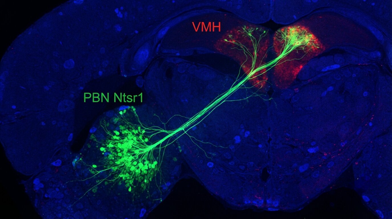
The population that senior author Richard Palmiter and his colleagues focused on expresses a gene called Ntsr1, which codes for a receptor for the neuropeptide neurotensin. These neurons, referred to throughout the study as PBN Ntsr1 neurons, form a small and anatomically scattered group within the lateral parabrachial nucleus. What makes them distinctive is their projection target: they send their axons almost exclusively to the ventromedial hypothalamus, a region known to be involved in both feeding behavior and the fear response. No other parabrachial population studied to date shows this combination of Ntsr1 expression and selective VMH projection.
To test what these neurons do, the researchers used chemogenetics, a technique in which neurons are engineered to express a designer receptor that can be activated by a specific synthetic drug that has no other biological effects. When they administered the drug to activate PBN Ntsr1 neurons, two things happened simultaneously. The animals reduced how much they ate, even after a period of fasting when hunger would normally drive robust food consumption. And they showed increased anxiety-like behavior on standardized behavioral tests designed to measure the willingness to explore potentially threatening environments.
The silencing experiment
To confirm that the neurons were not simply capable of producing both effects when artificially activated, but were actually necessary for normal anxiety-appetite interactions, the researchers performed the complementary experiment: they silenced the PBN Ntsr1 neurons permanently by expressing tetanus toxin light chain, a protein that blocks synaptic transmission, specifically in these cells. Animals in which PBN Ntsr1 neurons had been silenced showed the opposite pattern. They ate more. They were less anxious.
This bidirectional result is important for establishing the circuit’s normal function. Turning the neurons on produced anxiety and appetite suppression. Turning them off produced calm and increased appetite. The two outcomes moved together in both directions, suggesting that these neurons are not merely capable of linking the two systems under artificial stimulation, but are genuinely part of the circuitry that coordinates them under normal conditions.
What the neurons are doing in real time
The most revealing experiment in the study used fiber photometry, a technique that measures the fluorescence of calcium indicators in neurons in real time, allowing researchers to observe when the PBN Ntsr1 neurons are active during natural behavior. The researchers recorded from these neurons as animals navigated a variety of situations: eating freely, eating in environments designed to create anxious hesitation about approaching food, and encountering threatening stimuli.
The pattern that emerged was clean and consistent. The neurons fired more when animals were in threatening or anxiogenic situations. The same neurons went quiet when animals were actively eating. The temporal relationship between threat, neuronal activity, and eating was exactly what the circuit model predicted: danger activates these neurons, their activation suppresses appetite, and the relaxation of threat allows the neurons to quiet down and appetite to return.
This real-time recording established not just that the neurons could produce the effects when artificially triggered, but that they are actually recruited by threatening environments in the normal course of behavior and that their activity pattern corresponds to the behavioral suppression of feeding that threat produces.
The hypothalamic connection
The downstream target of PBN Ntsr1 neurons, the ventromedial hypothalamus, provides a plausible mechanism for how the circuit produces both anxiety and appetite suppression from a single neuronal population. The VMH has been known for decades to be involved in both feeding regulation and defensive behavior. Early lesion studies showed that damage to the VMH produced overeating and obesity, while electrical stimulation of the VMH produced fear and defensive responses. The region contains populations of neurons involved in satiety signaling and neurons involved in threat-related behavioral responses, and it receives inputs from multiple brain areas involved in both systems.
When PBN Ntsr1 neurons activate specific VMH populations, they appear to engage the region’s dual role simultaneously. The SF1 and BDNF-expressing neurons in the VMH that receive direct input from PBN Ntsr1 neurons are implicated in both energy balance and defensive behavior. The convergence of these functions in the VMH, and the convergence of the parabrachial input that drives both, suggests that the circuit identified in this study represents a specific implementation of the VMH’s known role in coordinating threat response and appetite suppression.
What this means beyond mice
The PBN Ntsr1 neurons and the circuit they form are identified in mice, and the study does not directly test whether an equivalent population exists in humans or whether it functions in the same way. The parabrachial nucleus is a conserved structure across mammalian species, and Ntsr1-expressing neurons in this region have been identified in other species, but the specific connectivity and functional role identified here will require human neuroscience tools, including neuroimaging and potentially direct recordings in clinical contexts, to verify in people.
The behavioral profile produced by activating these neurons — anxiety that suppresses appetite, particularly in environments where eating requires moving into open or exposed spaces — closely resembles the clinical presentation of anxiety-related eating disorders, in which threat hypersensitivity produces chronic suppression of appetite and avoidance of eating in social or public contexts. Whether PBN Ntsr1 neuron activity contributes to this clinical pattern in humans is a question the study opens but cannot yet answer.
What the study establishes is a mechanistic link between threat detection and appetite suppression that operates through a defined, genetically identifiable circuit rather than through diffuse neurochemical effects that are difficult to target specifically. A circuit with a specific cellular identity and a specific projection is also a circuit that can, in principle, be targeted by interventions designed to modulate it selectively — without the broad effects that come from targeting entire neurotransmitter systems or brain regions that serve multiple functions simultaneously.
Source
Jordan L. Pauli, Sekun Park, Rachel R. Felix, Richard D. Palmiter. “Parabrachial Ntsr1 neurons modulate food intake and anxiety through a projection to the ventromedial hypothalamus.” Proceedings of the National Academy of Sciences, 2026.
DOI: 10.1073/pnas.2605466123