People with narcolepsy collapse when they laugh or feel joy. Scientists just found the brain circuit responsible, and it runs through the love hormone

Narcolepsy is one of the most misunderstood neurological conditions in existence. Most people who have heard of it imagine someone falling asleep unexpectedly — a comedic image of a person dozing off mid-sentence or slumping over a desk. The reality is considerably more complicated, and for the approximately three million people worldwide who live with the condition, considerably less funny. One of its defining features is cataplexy: sudden episodes of complete or partial muscle paralysis that strike without warning and leave the person fully conscious but physically unable to move. These episodes can last from a few seconds to several minutes. They are not triggered by fatigue. They are triggered by happiness.
A person with narcolepsy is most likely to experience muscle paralysis when they laugh, when they reunite with someone they love, when they hear good news, or when they eat something they find delicious. For decades this pattern has been known and studied without a satisfying neurological explanation. Why would the brain collapse on itself at the exact moment it is feeling its best? A new study from Harvard Medical School, published in Nature Neuroscience, has now mapped the molecular and circuit-level answer. And it involves a molecule most people associate with love and bonding rather than paralysis.
What oxytocin is doing in the narcoleptic brain
Oxytocin is sometimes called the love hormone or the bonding neuropeptide. It surges during social interactions, physical touch, and rewarding experiences. It promotes trust, attachment, and the subjective sense of warmth that accompanies positive human connection. Its release is triggered by exactly the kinds of moments that also trigger cataplexy in people with narcolepsy: seeing friends, laughing, experiencing pleasure.
This parallel was not lost on Carrie Mahoney and her colleagues at Beth Israel Deaconess Medical Center, who set out to test whether the same neurochemical signal that makes social moments feel good might also be the signal that, in narcoleptic brains, causes those moments to go catastrophically wrong at the motor level.
They worked with a mouse model of narcolepsy whose orexin neurons have been genetically deleted. Orexin, also known as hypocretin, is the neuropeptide whose loss causes narcolepsy in humans and in these mice. Without orexin, the arousal system loses a critical stabilizing influence, and the boundary between wakefulness and the muscle paralysis of REM sleep becomes unstable. In affected mice, as in affected people, positive social and rewarding stimuli can tip that unstable boundary into collapse.
The team designed a test called the Social Isolation Reunification Test. Narcoleptic mice were briefly separated from their littermates, then reunited. The reunion triggered a sharp increase in cataplexy episodes — abrupt behavioral arrests lasting 30 to 60 seconds during which the mouse was fully conscious but unable to move. When the researchers administered an oxytocin antagonist, a drug that blocks oxytocin receptors, those socially triggered cataplexy episodes were eliminated. The mice still experienced cataplexy at baseline levels. The specifically social component of their cataplexy disappeared.
The moment before collapse
To understand what was happening at the level of individual neurons, the researchers used fiber photometry to record the activity of oxytocin-responsive neurons in the central amygdala in real time. The central amygdala is a brain structure involved in processing emotional salience, fear, reward, and the motivational significance of sensory experiences. It is connected to both the systems that generate positive social emotions and to the brainstem circuits that control muscle tone.
What the recordings showed was a precise and reliable sequence of events in the seconds before each cataplexy episode. Oxytocin levels in the central amygdala increased in the moments just before the mice collapsed. Simultaneously, the activity of neurons in the central amygdala that express oxytocin receptors — a population the researchers designated CeAOTR neurons — ramped upward. The neurons were most active just before the onset of paralysis, not during it.
This temporal sequence established the causal direction. Oxytocin arriving in the central amygdala was activating specific neurons there, and those neurons were somehow triggering the muscle paralysis that followed. To confirm this, the researchers used chemogenetics to artificially activate the CeAOTR neurons. Mice whose CeAOTR neurons were activated by a designer drug showed dramatically increased cataplexy. Mice whose CeAOTR neurons were chemically silenced showed dramatically reduced cataplexy, including reduced cataplexy during social reunification.
Optogenetics — the use of light-activated proteins to control neuron firing with millisecond precision — produced the same results even more cleanly. Shining light onto CeAOTR neuron cell bodies triggered cataplexy almost immediately. Shining light onto the axon terminals of those neurons at their downstream target in the brainstem did the same.
The circuit from love to paralysis
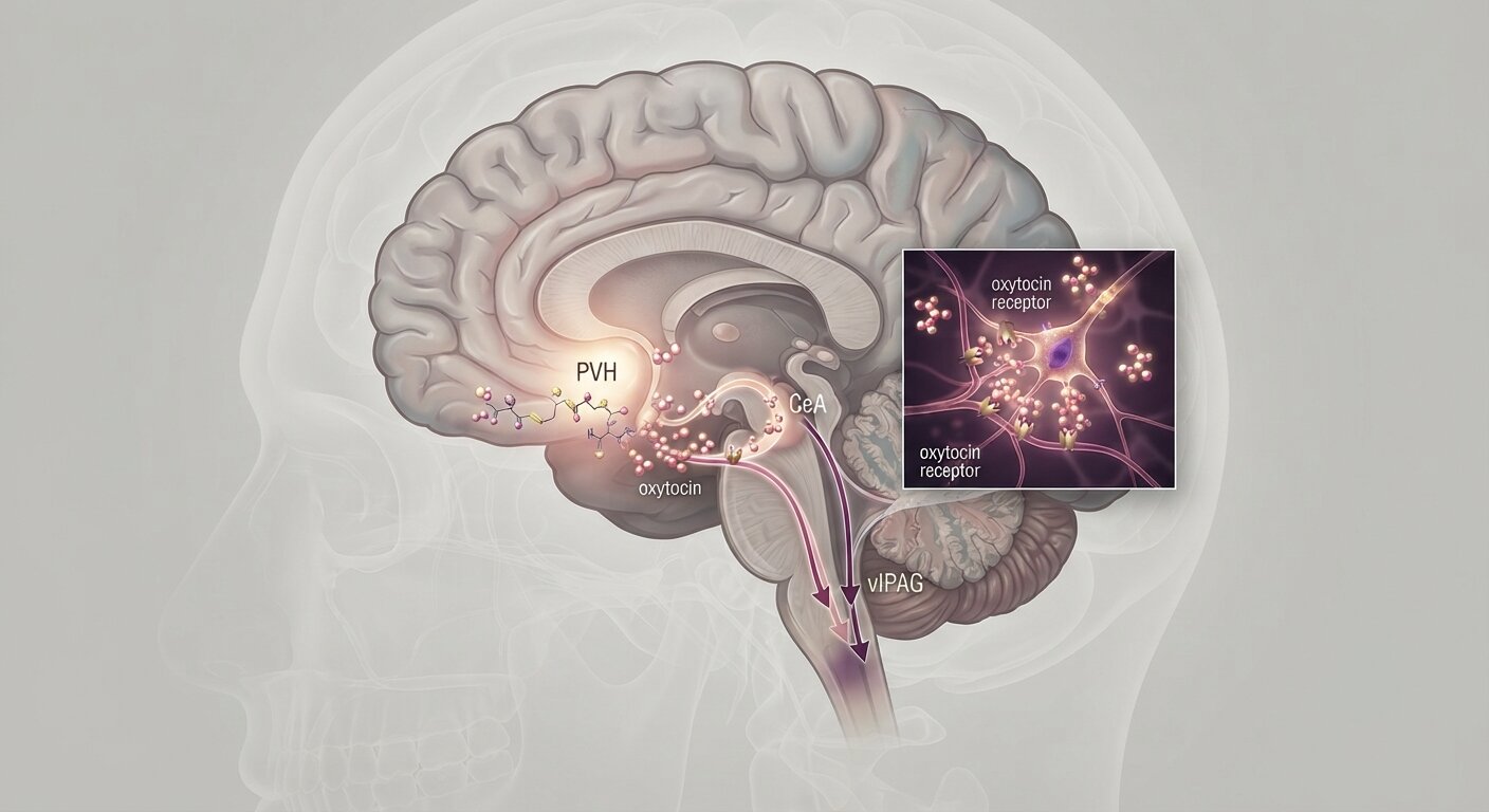
The full pathway the study documents is now mapped from beginning to end. When a person or narcoleptic mouse experiences a socially or emotionally rewarding moment, oxytocin is released from a region called the paraventricular nucleus of the hypothalamus. It travels to the central amygdala and activates CeAOTR neurons there. Those neurons then send inhibitory signals to a region called the ventrolateral periaqueductal gray, or vlPAG, which is a critical hub in the brainstem for maintaining muscle tone during wakefulness. The vlPAG normally suppresses the neural circuits responsible for the muscle atonia of REM sleep — the state in which the body is deliberately paralyzed to prevent us from acting out our dreams.
When CeAOTR neurons inhibit the vlPAG, its suppression of muscle atonia weakens. The REM sleep paralysis circuit is no longer being held fully in check. If the orexin system is intact, as it is in healthy people, orexin can compensate for this and maintain muscle tone regardless of what the CeAOTR neurons are doing. But in narcolepsy, where orexin neurons have been lost, that compensatory signal is absent. The CeAOTR neurons therefore have an outsized and unchecked effect. The result is that the most socially pleasurable and emotionally joyful moments of the day become the moments most likely to produce sudden muscle paralysis.
The system appears to have evolved to link positive social experiences to a mild relaxation of motor tone — a kind of physiological correlate of letting your guard down in the presence of trusted others. In people without narcolepsy, orexin keeps this relaxation within safe limits. In people with narcolepsy, orexin cannot do its job, and what should be a slight loosening of muscle tone becomes a complete collapse of it.
Chocolate and the same circuit
One of the more vivid details of the study concerns chocolate. It has long been known anecdotally and in research contexts that people with narcolepsy often report cataplexy triggered by eating chocolate or other strongly pleasurable foods. In mice, a similar phenomenon has been documented with chocolate exposure. The present study confirmed this and showed that chocolate-triggered cataplexy uses the same oxytocin-amygdala circuit identified for social triggers.
When narcoleptic mice were given access to chocolate, CeAOTR neurons became active — in the same way and with the same time course that they did during social reunification. Blocking the CeAOTR neurons pharmacologically or genetically reduced chocolate-induced cataplexy just as it reduced socially induced cataplexy. The pathway from positive emotion to muscle paralysis appears to be general: it responds to rewarding stimuli of multiple types, not only social ones, and it does so via the same molecular relay.
What blocking oxytocin could mean for treatment
The current treatments for cataplexy are imperfect. Sodium oxybate, the most effective medication available, requires taking doses in the middle of the night and has significant side effect profiles. Antidepressants suppress cataplexy somewhat by blocking the release of various neurotransmitters, but their effects are partial and come with the full array of antidepressant side effects.
An oxytocin antagonist that could block cataplexy specifically, without affecting the broader sleep-wake architecture, would represent a substantially different therapeutic approach. The experiments in this study showed that blocking oxytocin receptors eliminated the socially triggered component of cataplexy without measurably changing the amounts of time mice spent in wakefulness, NREM sleep, or REM sleep. The baseline rate of non-socially-triggered cataplexy was also not significantly affected.
This selectivity is clinically meaningful. A drug that targets specifically the social and reward-triggered pathways to cataplexy would allow patients to reduce their most situationally disruptive episodes — the ones that prevent them from laughing at a joke with friends, from experiencing joy at a reunion, from eating a meal they enjoy — without the systemic effects of current approaches.
Oxytocin antagonists already exist as licensed medications. Atosiban is approved for use in preterm labor in Europe. Other compounds with different pharmacokinetic profiles have been developed and tested in humans for various indications. The existence of clinically validated oxytocin antagonists means the path from this mechanistic finding to a therapeutic application is considerably shorter than it would be if the target were novel.
The researchers are careful to note that the work has been conducted in mice, and that the translation from mouse circuit-level findings to human therapeutic benefit is never guaranteed. The orexin knockout mouse model produces cataplexy through a mechanism that closely parallels human narcolepsy, but the precise oxytocin dynamics in human cataplexy will need to be verified directly. Whether the social and reward triggers of human cataplexy involve the same CeAOTR circuit will require human neuroimaging studies during cataplexy episodes, which are technically challenging given that cataplexy is brief, variable, and unpredictable.
What the study provides — the first mechanistic account of why positive social and emotional stimuli specifically trigger cataplexy in narcolepsy, mapped to the level of individual neuron populations and confirmed through multiple independent experimental methods — is the most complete answer the field has produced to a question narcolepsy patients have been asking for decades. Why does joy make the body collapse? Because the same hormone that signals happiness to the brain also tells the muscles, in a narcoleptic context, that it is safe to let go.
Source
Carrie E. Mahoney, Roberto De Luca, Adam A. Joyal, Caroline Woods, Wenling Zhao, Alissa A. Coffey, Emi Kurimoto, Daniel Kroeger, Lin Zhu, Henning Fenselau, Valery Grinevich, Christian R. Burgess, Elda Arrigoni, Thomas E. Scammell. “Oxytocin promotes socially triggered cataplexy.” Nature Neuroscience, 2026.
DOI: 10.1038/s41593-026-02352-7